Microscopes have revolutionized science and medicine by allowing us to observe objects and organisms that are invisible to the eye.
From the humble beginnings of simple lenses to the sophisticated electron microscopes of today, the ability to magnify and resolve the minutest details has expanded our understanding of the microscopic world.
This article explores the limits of microscopy, examining how small a microscope can really see and how many times an object can be magnified.
Historical Context and Basic Optical Microscopy
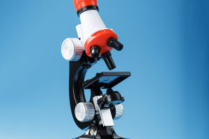
The journey of microscopy began in the late 16th century with the invention of the optical microscope. Early microscopes, such as those used by Antonie van Leeuwenhoek, could magnify objects up to about 200 times (200x). These simple light microscopes revealed a world teeming with microorganisms, previously unknown to humanity. Modern optical microscopes have significantly improved, offering magnifications up to 1,000 times (1,000x) and resolutions of about 200 nanometers.
Optical microscopes function by using visible light to illuminate the sample, with glass lenses magnifying the image. The resolution of these microscopes is fundamentally limited by the wavelength of visible light, which ranges from approximately 400 to 700 nanometers. As a result, structures smaller than this range cannot be resolved clearly with optical microscopes alone.
Breaking the Limits: Electron Microscopy
To see even smaller structures, scientists developed electron microscopes in the 20th century. These instruments use beams of electrons instead of light, which have much shorter wavelengths (down to fractions of a nanometer), allowing for far greater resolution.
Transmission Electron Microscopes (TEM): TEMs can magnify objects up to 10 million times (10,000,000x) with a resolution of about 0.1 nanometers. This allows for the observation of individual atoms under optimal conditions. TEMs work by transmitting electrons through a very thin specimen. The electrons interact with the specimen, and an image is formed based on how the electrons are scattered.
Scanning Electron Microscopes (SEM): SEMs provide detailed three-dimensional images of the surface of specimens with magnifications up to 500,000 times (500,000x) and resolutions of about 1 nanometer. SEMs work by scanning a focused beam of electrons across the specimen's surface. The electrons interact with the atoms on the surface, producing signals that can be used to form a detailed image.
Scanning Probe Microscopy
Another breakthrough in microscopy is scanning probe microscopy, which includes techniques such as atomic force microscopy (AFM) and scanning tunneling microscopy (STM). These methods do not use lenses or electron beams but instead scan a physical probe over the surface of the sample.
Atomic Force Microscopy (AFM): AFMs can achieve resolutions on the order of fractions of a nanometer. An AFM uses a cantilever with a sharp tip that interacts with the sample surface at the atomic level. As the tip scans the surface, it deflects based on the forces between the tip and the sample, creating a high-resolution image of the surface topology.
Scanning Tunneling Microscopy (STM): STMs also achieve atomic resolution. They work by bringing a conductive tip extremely close to the surface of a sample, allowing electrons to "tunnel" through the vacuum between the tip and the sample. This tunneling current is measured and used to construct an image of the surface at atomic resolution.